Introduction
Hypertension remains one of “the most significant non-infection pandemiae in human history contributing to a pattern of cardiovasular morbidity and mortality” [1]. Hypertension contributes to one out of every seven deaths in the United States, and approximately 70% of persons who have a first heart attack or stroke are hypertensive. Prevalence of the arterial hypertension increases with age to approximately 70% among persons aged 65 years and older [2]. It’s necessary to note that medical tactics and cardiovascular risk stratification in patients with arterial hypertension strongly depend on type and intensity of target organs injury and associated comorbidity.
Vascular wall is a well-known target organ in hypertension. High-resolution ultrasound imaging of arteries (USIA), a convenient noninvasive method for evaluating carotid arterial walls, became a standard in clinical investigation of hypertensive patients [3]. USIA depicts 2 main clinical markers of vascular lesion: increased intima media thickness (IMT) and plaque formation. Both changes are related to ageing and are accelerated by hypertension and other risk factors. However, the majority of clinical trials have suggested that those findings were affected by different cardiovascular risk factors.
With regard to relation to ageing, some previous studies had indicated that IMT as well as atherosclerotic plaque formation are both age dependent processes, which aggravation is due to hypertension and other cardiovascular risk factors [4, 5]. But the results of the majority of clinical trials had been received in adult population (patients older than 65 were mostly excluded), on the other hand the very involutional process influence upon vascular wall remodeling remains in the area of uncertainty. All mentioned above encouraged us to study age dependent vascular remodeling in elderly patients with arterial hypertension.
Material and Methods
179 elderly and senile patients with arterial hypertension were included in the study. Exclusion criteria were the following: stroke and transient ischemic attacks in 12 months and less prior involvement in the study; thyroid pathology; endocrine disorders with hypertension; usage of drugs with systemic hypertensive effects; renal disorders preceding with blood pressure (BP) increase (according to medical documentation); central hypertension; cognitive deficit impairing clinical investigation, arrhythmias (with exception of non-frequent extrasystolia); chronic heart failure of functional classes III and IV (NYHA); angina of classes III and IV (Canadian cardiovascular society classification); acute coronary event in 6 months or less prior to involvement in the study; diabetes mellitus; glomerular filtration rate less than 60 ml/min/1.73 m2; oncologic and haematologic disorders; comorbidity exacerbation.
Intima media thickness was assessed by means of ultrasound system Philips Envisor HD (USA) according to a Consensus Statement from the American Society of Echocardiography Carotid Intima-Media Thickness Task Force Endorsed by the Society for Vascular Medicine (2008). Both right and left carotid arteries were studied.
Target blood pressure levels were considered as according to national guideline on diagnostic and treatment of the arterial hypertension [1]. In comparison analysis stable target blood pressure criteria was calculated as the ratio of all cases of elevated blood pressure episodes and number of days of observation. The only strict condition of this criteria calculation was continuous blood pressure self-control and antihypertensive medication.
Received data were put together into the analytical data base. Certain parameters were indexed while several parameters were ranged according to the study protocol. Normality was checked by Shapiro-Wilks criteria, skewness and kurtosis calculation. Statistical hypothesis were checked by 2-sided Mann-Whitney test. Correlation analysis by Spearmen, Kendall and gamma correlation were performed. Basing on presumed type of time function, polynomial approximation of received parameters’ values by means of least squares method was performed. Established time functions were used to construct first derivatives and phase patterns on “time function – it’s first derivative” surface. Data are presented as mean±SD unless otherwise indicated. Probability level (p) less than 0.05 was considered statistically significant.
Results
Mean age of the patients included in the study was 68,44 years (age range was 60-84 years). In 62.01% of patients included into the study age was in the range of 60-74 (age group I) while in 37.99% it was in the range of 75 – 89 (age group II). 54.19% of studied patients were male, and 45.81% were female.
Ageing was associated with statistically significant increase in IMT (Table 1). Intima media thickness was significantly larger in male patients: 0.10±0.01 cm vs 0.09±0.02 cm, 2-sided Man-Whitney test p level 0.045 – right common carotid artery, and 0.11±0.02 cm vs 0.09±0.01 cm, 2-sided Mann-Whitney test p level 0.004 – left common carotid artery (Table 2). Comparison of elderly and senile groups of patients revealed similarity in sex pattern, clinical signs and symptoms frequencies, screening biochemical serum parameters, medication and blood test parameters. This fact allowed us to use combined groups in further going analysis. No significant differences between patients with controlled and uncontrolled hypertension were established (Table 3).
Table 1. Morphological parameters of CCA in elderly patients with hypertension: comparison of age groups
|
|
Age group I |
Age group II |
|
Diameter CCA R, cm, mean±SD |
0.75±0,08 |
0.74±0,06 |
|
Diameter CCA L, cm, mean±SD |
0.72±0,15 |
0.77±0,04 |
|
Kinking CCA R, mode (frequency of mode) |
0 (97) |
0 (12) |
|
Kinking CCA L, mode (frequency of mode) |
0 (101)* |
0 (9)* |
|
IMT CCA R, cm, mean±SD |
0.07±0,04* |
0.09±0,04* |
|
IMT CCA L, cm, mean±SD |
0.12±0,11* |
0.14±0,02* |
CCA – common carotid artery; R – right; L – left; * - significant differences.
Table 2. Morphological parameters of CCA in elderly patients with hypertension: gender comparison
|
|
Male |
Female |
|
CCA R Diameter, cm, mean±SD |
0.76±0,08* |
0.73±0,07* |
|
Diameter CCA L, cm, mean±SD |
0.77±0,08* |
0.69±0,17* |
|
Kinking CCA R, mode (frequency of mode) |
0 (57) |
0 (52) |
|
Kinking CCA L, mode (frequency of mode) |
0 (54) |
0 (56) |
|
IMT CCA R, cm, mean±SD |
0.08±0,04 |
0.07±0,04 |
|
IMT CCA L, cm, mean±SD |
0.13±0,14* |
0.11±0,02* |
CCA – common carotid artery; R – right; L – left ; * - significant differences.
Table 3. Morphological parameters of CCa in elderly patients: comparison of controlled and non-controlled hypertensive patients
|
|
Controlled |
Non controlled |
|
Diameter CCA R, cm, mean±SD |
0.75±0,08* |
0.74±0,07* |
|
Diameter CCA L, cm, mean±SD |
0.74±0,11* |
0.67±0,22* |
|
Kinking CCA R, mode (frequency of mode) |
0 (80)* |
0 (21)* |
|
Kinking CCA L, mode (frequency of mode) |
0 (80)* |
0 (23)* |
|
IMT CCA R, cm, mean±SD |
0.07±0,04* |
0.06±0,04* |
|
IMT CCA L, cm, mean±SD |
0.11±0,08* |
0.14±0,16* |
CCA – common carotid artery; R – right; L – left.
* - non significant differences.
Age dependent intima media thickness was of nonlinear character, and remained rather stable up to age of 74 years. After that age, briskly augmentation of the IMT with mean rate equal to 0.157 cm/year occurred (Fig. 1). Phase pattern analysis revealed stable focuses (0.114; 0) – for left carotid artery, and (0.105;0) for right carotid artery (Fig. 2).
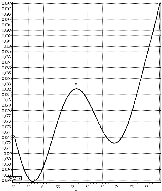
Figure 1. IMT CCA R time function

Figure 2. Phase pattern of cerebrovascular system on “IMT (CCA R) time function – IMT (CCA R) rate” surface
Atherosclerotic plaque detection frequency significantly increased from 7th decade of life duration. No significant correlations within IMT and systolic, diastolic, pulse, mean blood pressure levels, as well as within IMT and screening biochemical and haematological parameters were detected (Table 4). IMT was associated with granulocytes and lymphocytes counts: (r=0.395; 0.319, p level <0.05; and r=-0.323; 0.326 p level<0.05, for right and left common carotid arteries relatively) (Table 4).
Table 4. Correlation matrix (Spearman R)
|
|
IMT CCA R |
IMT CCA L |
|
SBP, standing |
-0.024898 |
0.119210 |
|
DBP standing |
0.111652 |
0.178477 |
|
MMI LV |
0.271860 |
0.082047 |
|
Cholesterol |
0.122906 |
0.184423 |
|
Creatinin |
0.128615 |
0.212722 |
|
Erythrocytes |
-0.116087 |
-0.022055 |
|
Hb |
-0.100464 |
-0.015791 |
|
Platelets count |
0.030438 |
0.017567 |
|
Leukocytes count |
0.255655 |
0.181109 |
|
Granulocytes count |
0.395318* |
0.319727* |
|
Lymphocytes count |
-0.323199* |
-0.326141* |
|
Monocytes count |
-0.101382 |
0.002922 |
|
HDLP |
0.318830 |
0.049031 |
|
LDLP |
0.296372 |
0.210614 |
|
TG |
0.226499 |
0.050482 |
|
MCV |
-0.062272 |
0.011615 |
|
MCH |
-0.040641 |
-0.011703 |
|
RDW |
0.151228 |
0.090151 |
|
MPV |
-0.114717 |
-0.044284 |
|
PDW |
-0.019324 |
0.023633 |
|
Ht |
0.056775 |
0.077729 |
SBP – systolic blood pressure, DBP – diastolic blood pressure, MMI – myocardial mass index; Hb – hemoglobin concentration; HDLP – high density lipoproteins; LDLP – low density lipoproteins; TG – triglycerides; MCV – mean cell volume of erythrocytes; MCH – mean hemoglobin concentration in erythrocytes; RDW – red blood cell distribution width; MPV – mean platelets volume; PDW – platelets distribution width; Ht – hematocrit, * - p level < 0.05.
Discussion
Nowadays IMT dependence upon age is beyond any doubts, but the characteristics of this dynamics as well as intimate mechanism this augmentation remains unclear. In several studies of IMT in several age groups all measurements were performed without taking atherosclerotic lesion into account [6, 7]. In means that data were received by measurements in full range from plaque free to significant atherosclerotic lesion zones of carotid arteries. Exclusion criteria were not strict enough in other trials, for example patients with diabetes mellitus and peripheral artery disease were not excluded from the study.
In our study all measurements were performed at unaffected areas of vascular wall. We tried to use strict criteria to avoid all possible vasculopathies as well as symptomatic secondary genesis of arterial hypertension in patients involved in the study. Patients of age groups I and II were totally comparable by gender and clinical characteristics, and medication spectrum.
There are lots of studies suggesting existence of correlation between IMT and blood pressure volumes. For instance, paper of di Bello (2009) postulated significant correlation between pulse pressure and IMT, but this data are no doubt questionable, for r value in this work was equal to only 0.19, and no analysis of nonlinear correlation was mentioned. In our study no data sustained possibility of blood pressure level influence upon IMT was received [8]. So, the possible conclusion is that age dependent increase of IMT belongs to involutional process in human arteries.
Characteristics of age dependent IMT dynamics are also controversial. In majority of studies nonlinear type was detected. In the same time S. Homma and coauthors had studied practically healthy persons in age range from 21 up to 105 years old and revealed linear IMT dependence on age [9]. The increase in IMT reflects predominantly intimal thickening. When sites with plaques are used to measure IMT, progression of IMT will show exponential acceleration in the elderly. In contrast, mean IMT in plaque-free sites reveals nonlinear association with age.
Taking into consideration deciphered mechanisms of vascular remodeling, linear age dependent increase of IMT seems to be hardly probable [10, 11, 12]. According to biomechanics, the normal artery is subjected to 3 primary mechanical stresses: a blood flow-induced wall shear stress, a blood pressure-induced circumferential wall stress, and an axial wall stress that appears to arise during development and to persist into maturity because of the long half-life of elastin. Coupling of mechanic stresses and biomechanical vessel wall characteristics influence can mostly explain oscillations of IMT during ageing in studied sample. Hence, further investigations of intimate age dependent vascular remodeling process are strongly necessary.
The majority of the IMT studies postulates association between IMT and atherosclerotic plaque formation. Contrary to that in our study such association was not revealed, and this fact also indicated speciality of vascular remodeling in elderly and senile patients.
Conclusion
In elderly patients age dependent diffuse increase of intima media thickness was detected. The dynamics of vascular remodeling was of nonlinear character, and was independent on degree and duration of hypertension. Received data can be considered as a reasonable doubt of intima media thickness usage as atherosclerotic lesion markers in elderly patients.
Conflict of interest: Nothing to declare.
- Prophylactics, diagnosis and treatment of arterial hypertension in Russian Federation (experts position paper). RMJ 2000; 8: 318-349 [rus].
- Yoon PW, Gillespie CD, George MG, Wall HK. Control of hypertension among adults - national health and nutrition examination survey, United States, 2005-2008. MMWR Surveill Summ 2012. 61(2): 19-25.
- Diagnostics and treatment of arterial hypertension: national guidelines / ed. R.G. Oganov, 3d edition. Moscow: Siliceya Poligraph, 2010: 464-500 [rus].
- Espeland MA, Hoen H, Byington R, Howard G, Riley WA, Furberg CD. Spatial distribution of carotid intimal-medial thickness as measured by B-mode ultrasonography. Stroke 1994; 25(9):1812-1819 (doi: 10.1161/01.STR.25.9.1812).
- Bonithon-Kopp C, Raison J, Courbon D, Bonhomme G, Guy-Grand B, Ducimetière P. Relation of intima-media thickness to atherosclerotic plaques in carotid arteries: the vascular aging (EVA) study. Arterioscler Thromb Vasc Biol 1996; 16(3): 310-316 (PMID: 8620348) (doi: 10.1161/01.ATV.16.2.310).
- Salonen R, Salonen JT. Progression of carotid atherosclerosis and its determinants: a population-based ultrasonography study. Atherosclerosis 1990. 81: 33-40 (PMID: 2407252).
- Allan PL, Mowbray PI, Lee AJ, Fowkes FG. Relationship between carotid intima-media thickness and symptomatic and asymptomatic peripheral arterial disease: The Edinburgh Artery Study. Stroke 1997; 28(2): 348-353 (PMID: 9040688) (doi: 10.1161/01.STR.28.2.348).
- Di Bello V, Carerj S, Perticone F, Benedetto F, Palombo C, Talini E, Giannini D, La Carrubba S, Antonini-Canterin F, Di Salvo G, Bellieni G, Pezzano A, Romano MF, Balbarini A; Research Group of the Italian Society of CardioVascular Echocardiography (SIEC). Carotid intima-media thickness in asymptomatic patients with arterial hypertension without clinical cardiovascular disease: relation with left ventricular geometry and mass and coexisting risk factors. Angiology 2009; 60(6): 705-713 (doi: 10.1177/0003319708329337).
- Homma S, Hirose N, Ishida H, Ishii Y, T. Araki G. Carotid Plaque and Intima-Media Thickness Assessed by B-Modeltrasonography in Subjects Ranging From Young Adults to Centenarians. Stroke 2001; 32(4): 830-835 (doi: 10.1161/01.STR.32.4.830).
- Jamaluddin MS, Weakley SM, Zhang L, Kougias P, Lin PH, Yao Q, Chen C. miRNAs: roles and clinical applications in vascular disease. Expert Rev Mol Diagn 2011; 11(1): 79-89 (doi:10.1586/erm.10.103) (PMID: 21171923).
- Duprez DA. Systolic hypertension in the elderly: addressing an unmet need. Amer J Med 2008; 121(3): 179-184. e3 (PMID: 18328297) (doi:10.1016/j.amjmed.2007.10.027).
- Seals DR, Moreau KL, Gates PE, Eskurza I. Modulatory influences on ageing of the vasculature in healthy humans. Exp Gerontol 2006; 41(5): 501-507 (PMID: 16537099) (doi: 10.1016/j.exger.2006.01.001).
Received 15 May 2012, Accepted 10 June 2012.
© 2012, Malinova L.I., Sadjaya L.A., Tikhonova L.A.
© 2012, Russian Open Medical Journal
Author's edition of the text
