

Introduction
Urolithiasis is one of the most common diseases in contemporary urology. It poses an important global health problem [1-3]. The prevalence and incidence of urolithiasis have been steadily increasing in recent decades, and current estimates suggest that up to 10–15% of the adult population will experience urolithiasis at least once in their lifetime [4, 5]. Urolithiasis predominantly affects working-age individuals and is associated with significant healthcare costs due to recurrence, repeat interventions, and long-term follow-up [6, 7]. Despite advances in diagnosis and treatment, recurrence rates remain high, highlighting the need for improved strategies in both treatment and prevention [8, 9].
The clinical course of urolithiasis is highly heterogeneous and depends on a complex interplay of metabolic, environmental, and anatomical factors [10, 11]. Kidney stone composition plays a central role in determining its fragility, response to treatment, and risk of recurrence [12, 13]. Calcium oxalate stones remain the most common worldwide. However, the proportion of calcium phosphate, uric acid, and mixed stones varies significantly between populations [14]. These differences are not random and reflect regional variations in climate, elevation, water composition, dietary habits, and hydration status [15].
Minimally invasive surgical techniques have become the standard of care for urolithiasis, largely replacing open surgery [16]. Currently, extracorporeal shock wave lithotripsy (SWL), percutaneous nephrolithotomy (PCNL), and ureteroscopy (URS), including retrograde intrarenal surgery (RIRS), are widely employed depending on stone size, location, and patient-related factors [17]. Although international guidelines provide general recommendations, the optimal choice of treatment modality remains controversial, particularly for large, complex, or recurrent stones [18, 19]. In routine practice, the choice of treatment modality is often determined by stone size and anatomical features, while other potentially important prognostic factors are underutilized [20, 21].
Computed tomography (CT) has become the imaging modality of choice for the assessment of urolithiasis [22, 23]. In addition to stone detection and localization, non-contrast CT provides quantitative information on stone density, measured in Hounsfield units (HU) [24]. Numerous studies have demonstrated a relationship between CT density, stone composition, and treatment outcomes, particularly fragmentation efficiency and stone-free rates after SWL and endoscopic procedures [25-27]. Higher HU values are generally associated with increased stone hardness and reduced treatment success. However, CT-derived parameters are not always integrated into clinical decision-making algorithms [28].
Geographic and environmental factors represent an additional – often overlooked – aspect in urolithiasis research [29]. Climatic conditions, elevation, mineral content of drinking water, and regional dietary patterns influence urine composition and stone formation [15, 29]. Mountainous and high-elevation regions are characterized by unique environmental conditions, including lower ambient temperatures, altered hydration patterns, and specific geochemical properties of water sources [30]. These factors may influence not only the incidence of urolithiasis but also the composition and structural characteristics of kidney stones, thereby influencing treatment efficacy [31].
The Kyrgyz Republic provides a unique natural environment for studying these interactions. A significant share of the population lives in mid- and high-elevation regions, where environmental and climatic conditions differ markedly from lowland areas [32]. Limited regional data suggest that urolithiasis in the Kyrgyz Republic may be associated with geographic variability, including differences in stone composition and clinical presentation [33]. However, systematic comparative analyses that integrate geographic region, stone characteristics, and minimally invasive treatment outcomes remain limited.
Most published studies focused on individual predictors of treatment success, such as stone size or CT parameters, rather than on integrated comparative analyses across different geographic contexts [34]. Data from high-elevation regions remain particularly limited, despite the potential clinical significance of these factors for treatment selection and outcome prediction.
Therefore, the goal of this study was to evaluate the comparative efficacy of minimally invasive stone treatment methods among patients from different geographic regions of the Kyrgyz Republic, with a particular emphasis on stone composition and density determined by CT scan.
Material and Methods
Study design and setting
This multicenter retrospective comparative study was conducted in the Kyrgyz Republic. Patients were treated between 2020 and 2023 at three specialized urology centers in Bishkek: the Department of Urology at Kyrgyz State Medical Academy, Avicenna Medical Center, and the Center for Urology at the National Hospital the Kyrgyz Republic. The study compared stone characteristics and minimally invasive treatment outcomes in patients living in mid- and high-elevation areas. Mid-elevation areas were defined as areas at an altitude of 1,200 to 2,000 meters above sea level, whereas high-elevation areas were above 2,000 meters above sea level, according to the national geographic classification used in the Kyrgyz Republic. This study was conducted and presented in accordance with the STROBE guidelines for observational studies.
Study population and selection criteria
The study included adult patients aged 18–75 years who underwent elective minimally invasive surgical treatment for urolithiasis during the study period. All patients had radiographic confirmation of urolithiasis.
Patients were included in the study if they had preserved renal function and no signs of obstructive uropathy or congenital urinary tract anomalies. Exclusion criteria were age under 18 or above 75 years, advanced chronic kidney disease (stages 4-5), acute kidney injury, obstructive uropathies, or anatomical abnormalities of the urinary tract that could impact treatment choice or outcomes.
A total of 139 patients met the inclusion criteria and were included in the final analysis. Based on their region of residence, patients were divided into mid-elevation (n=88) and high-elevation (n=51) groups. Eligible patients were identified from institutional medical records and screened according to predefined inclusion and exclusion criteria.
Kidney stone characteristics
The location, size, and composition of the stones were recorded for each patient. Stone size was estimated using preoperative non-contrast CT and defined as the maximum diameter (mm) of the primary stone. In patients with multiple stones, the largest stone was used as the primary stone. Stone location was classified by anatomical site (kidney and/or ureter).
Stone composition was determined by laboratory analysis of the extracted stone material, if available, and classified according to the predominant mineral component. Stones were classified as calcium oxalate, calcium phosphate, uric acid, or mixed. If complete stone material was not available, the composition was recorded based on information documented in the medical record.
CT assessment and stone density
Preoperative non-contrast CT was performed as part of the routine examination of all included patients. Stone density was measured in HU by placing a region of interest within the stone on axial images, avoiding surrounding soft tissue and minimizing partial volume effects. For irregularly shaped stones, measurements were taken in the central portion to ensure representative attenuation values. In patients with multiple stones, the HU value was recorded for the primary stone. If repeated measurements were available, the mean HU values were used.
Treatment methods
The treatment method was selected according to standard clinical practice based on stone size, location, anatomical features, and patient-related factors. Extracorporeal SWL, PCNL, and URS with or without RIRS were employed when necessary. The initial treatment method chosen was recorded for each patient. Additional sessions or secondary procedures, whenever necessary, were documented.
Treatment outcome measures
The primary outcome was treatment efficacy, assessed by stone freedom after the initial procedure. Stone-free status was defined as the absence of residual fragments on follow-up imaging or the presence of clinically insignificant residual fragments according to standard clinical criteria.
Secondary outcomes included the need for additional treatment sessions or secondary procedures, as well as procedure-related complications. The method and timing of follow-up imaging were determined based on standard clinical practice at the participating centers.
Statistical analysis
Continuous variables were presented as mean ± standard deviation or median (interquartile range [IQR]), as appropriate. Categorical variables were presented as frequencies and percentages. Comparisons between groups were performed using the Student’s t-test or Mann-Whitney U test for continuous variables and the chi-squared test or Fisher’s exact test for categorical variables, as applicable.
Associations between stone characteristics, CT-derived stone density, geographic group, and treatment outcomes were examined using univariate and multivariate analyses where appropriate. All tests were two-sided, and p<0.05 was considered statistically significant. Given the retrospective study design and limited sample size in subgroups, calculations of preliminary sample size or power, as well as multivariate regression analyses, were not performed.
Results
Patient characteristics
The study included 139 patients with urolithiasis, divided by place of residence into predefined mid-elevation and high-elevation geographic regions. The study population was predominantly male (74.8%), with a mean age of 55.85 years. Baseline demographic characteristics, body mass index, lifestyle factors, and comorbidities did not differ statistically significantly between groups (Table 1).
Table 1. Baseline characteristics of the study population by geographic region
|
Parameter |
Mid-elevation regions (n=88) |
High-elevation regions (n=51) |
Total (n=139) |
p-value |
|
Age, years |
0.248 |
|||
|
20-29 |
4 (2.9%) |
3 (2.2%) |
7 (5.0%) |
|
|
30-39 |
8 (5.8%) |
5 (3.6%) |
13 (9.4%) |
|
|
40-49 |
9 (6.5%) |
10 (7.2%) |
19 (13.7%) |
|
|
50-59 |
17 (12.2%) |
8 (5.8%) |
25 (18.0%) |
|
|
60-69 |
45 (32.4%) |
18 (12.9%) |
63 (45.3%) |
|
|
70-75 |
5 (3.6%) |
7 (5.0%) |
12 (8.6%) |
|
|
BMI, kg/m² (mean±SD) |
24.6±4.11 |
24.12±4.6 |
24.54±4.22 |
0.322 |
|
Gender |
||||
|
Female |
24 (17.3%) |
11 (7.9%) |
35 (25.2%) |
0.178 |
|
Male |
64 (46.0%) |
40 (28.8%) |
104 (74.8%) |
|
|
Lifestyle factors |
||||
|
Alcohol consumption |
16 (11.5%) |
23 (16.5%) |
39 (28.1%) |
0.761 |
|
Smoking |
19 (13.7%) |
39 (28.1%) |
58 (41.7%) |
0.058 |
|
Comorbidities |
||||
|
Diabetes mellitus |
7 (5.0%) |
7 (5.0%) |
14 (10.1%) |
0.567 |
|
Hypertension |
23 (16.5%) |
26 (18.7%) |
39 (35.3%) |
0.963 |
Stone characteristics
Stone location, laterality, and size are summarized in Table 2. Renal stones of various intrarenal locations were predominant in the study population.
Table 2. Stone characteristics by geographic region
|
Parameter |
Mid-elevation regions (n=88) |
High-elevation regions (n=51) |
Total (n=139) |
p-value |
|
Stone laterality |
0.270 |
|||
|
Right |
36 (25.9%) |
15 (10.8%) |
51 (36.7%) |
|
|
Left |
48 (34.5%) |
34 (24.5%) |
69 (59.0%) |
|
|
Bilateral |
4 (2.9%) |
2 (1.4%) |
6 (4.3%) |
|
|
Intrarenal location |
||||
|
Renal pelvis |
41 (29.5%) |
16 (11.5%) |
57 (41.0%) |
0.005 |
|
Ureteropelvic junction |
19 (13.7%) |
11 (7.9%) |
30 (21.6%) |
0.006 |
|
Lower calyx |
22 (15.8%) |
6 (4.3%) |
28 (20.1%) |
0.004 |
|
Middle calyx |
14 (10.1%) |
2 (1.4%) |
16 (11.5%) |
0.004 |
|
Upper calyx |
7 (5.0%) |
2 (1.4%) |
9 (6.5%) |
0.004 |
Stones were most often located on the left side (59.0%), while bilateral lesions were rare (4.3%). Stone laterality did not differ significantly between geographic groups.
In contrast, we observed statistically significant differences in intrarenal stone location. Stones located in the renal pelvis, ureterorenal junction, and lower and middle calyces were significantly more common in patients living in mid-elevation regions, compared with those living in high-elevation areas.
The distribution of stone sizes was similar between geographic groups. The most common size category was 1.5-2.0 cm (30.2%), followed by stones ≤1.5 cm (25.2%) and 2.1-3.0 cm (21.6%). Stones larger than 3.0 cm accounted for a smaller proportion of cases.
Stone composition by geographic region
Stone composition analysis revealed pronounced differences in the distribution of urinary stone types between geographic regions. The comparative distribution of stone composition among patients living in mid-elevation and high-elevation regions is shown in Table 3 and Figure 1.
Table 3. Stone composition by geographic region
|
Stone type |
Mid-elevation regions (n=88) |
High-elevation regions (n=51) |
Total (n=139) |
p-value |
|
Oxalate |
47 (53.4%) |
21 (41.2%) |
68 (48.7%) |
|
|
Phosphate |
23 (26.1%) |
16 (31.4%) |
39 (27.7%) |
|
|
Urate |
7 (8.0%) |
4 (7.8%) |
11 (7.6%) |
|
|
Mixed |
9 (10.2%) |
8 (15.7%) |
17 (12.6%) |
|
|
Cystine |
2 (2.3%) |
2 (3.9%) |
4 (3.4%) |
|
|
Overall comparison |
|
0.663 |
||
Data are presented as n (%). Intergroup comparisons were performed using the χ² test.
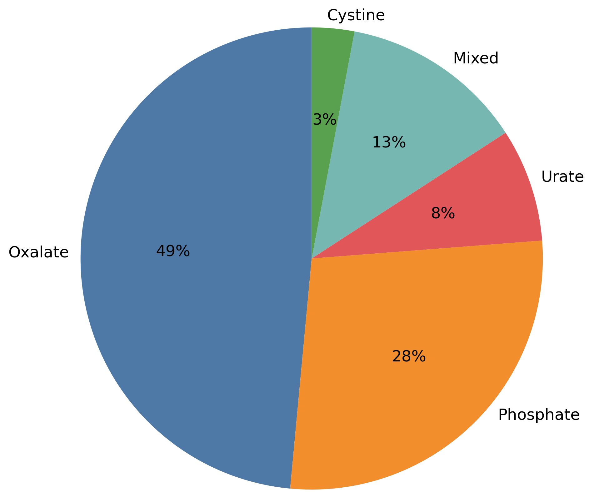
Figure 1. Distribution of urinary stone composition in the study population.
Oxalate and phosphate stones predominated in the overall study population, accounting for more than three-quarters of cases. Oxalate stones were more common among patients living in mid-elevation regions, while phosphate stones were relatively more common in high-elevation areas. Uric acid stones, mixed stones, and cystine stones were less common in both geographic groups.
Elemental composition of urinary stones by geographic region
The elemental composition of urinary stones was analyzed to assess potential differences related to geographic region. X-ray spectral analysis was performed on stone samples extracted during surgical procedures, allowing for the quantification of major inorganic oxides and trace elements. Comparative data for patients living in mid- and high-elevation regions are summarized in Table 4.
Table 4. Elemental composition of urinary stones by geographic region
|
Component (wt.%) * |
High-elevation regions |
Mid-elevation regions |
|
CaO |
30.0 |
30.0 |
|
MgO |
0.20 |
0.15 |
|
Na₂O |
0.50 |
0.50 |
|
SiO₂ |
0.30 |
0.30 |
|
Fe₂O₃ |
0.10 |
0.10 |
|
Al₂O₃ |
0.10 |
0.10 |
|
CuO |
0.005 |
0.008 |
|
PbO |
0.002 |
0.004 |
|
ZnO |
0.010 |
0.012 |
|
SrO |
0.015 |
0.018 |
Elemental analysis revealed that calcium oxide (CaO) was the predominant component of urinary stones in both geographic regions (median: 30.0 wt.% in each group), while sodium oxide (Na2O) levels were also similar (median: 0.50 wt.%). Stones from high-elevation regions had a higher median magnesium oxide (MgO) content (0.20 vs. 0.15 wt.%). Trace elements were present in low concentrations in both groups; median values of CuO, PbO, ZnO, and SrO were slightly higher in stones from mid-elevation regions. A relationship was found between calcium oxide content and stone density, as determined by CT, with higher CaO levels corresponding to higher HU values. A direct correlation was revealed between calcium oxide content and stone density determined by CT, indicating higher HU values with increasing CaO concentration (r=0.71, p<0.001; Figure 2).
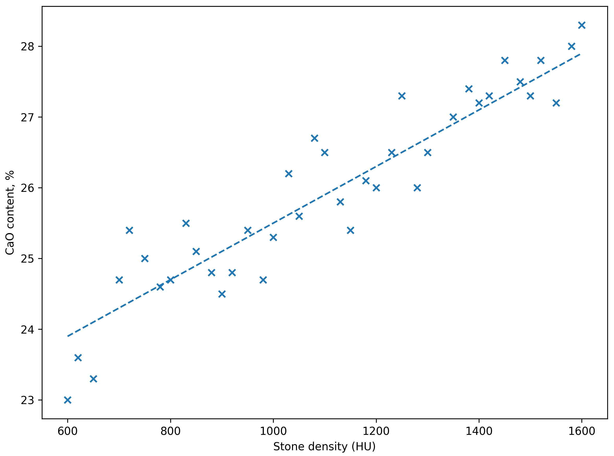
Figure 2. Relationship between calcium oxide (CaO) content and stone density (HU) determined by CT scan. Higher CaO content is associated with elevated stone density.
Treatment methods and outcomes
Minimally invasive treatment methods were selected according to standard clinical practice based on stone size, location, and anatomical features. Treatment approaches and outcomes were analyzed by geographic region and stone characteristics.
Treatment outcomes by treatment method and geographic region
Treatment outcomes were assessed by treatment method and compared between patients residing in mid- and high-elevation regions. The primary endpoint was stone freedom after initial minimally invasive intervention, assessed by postoperative imaging (Table 5).
Table 5. Treatment outcomes and resource indicators by method and geographic region
|
Treatment method |
High-elevation regions (n, %) |
Mid-elevation regions (n, %) |
p-value |
|
Percutaneous nephrolithotomy |
27/31 (87.1%) |
36/44 (81.8%) |
0.751 |
|
Ureteroscopy / RIRS |
17/19 (89.5%) |
27/31 (87.1%) |
1.000 |
|
Shock wave lithotripsy |
14/17 (82.4%) |
21/27 (77.8%) |
1.000 |
RIRS, retrograde intrarenal surgery. Stone freedom was defined as the absence of residual fragments >4 mm at follow-up, as determined by imaging. Intergroup comparisons were performed using the χ² test.
Stone freedom rates were high with PCNL and URS in both geographic regions. SWL demonstrated lower rates of stone freedom vs. endoscopic methods, with a trend toward reduced efficacy in patients residing in mid-elevation regions. No statistically significant differences in stone freedom rates were observed between geographic groups for any treatment modality.
Infectious complications were uncommon and did not exceed 10% across all treatment modalities. A higher proportion of infectious complications was observed among patients undergoing URS in mid-elevation regions vs. high-elevation regions, while complication rates for PCNL and SWL remained low and were similar in both geographic groups. No severe postoperative complications were reported.
Additional treatment or repeat intervention was required in some patients, most commonly after SWL and URS. Repeat procedures were more frequently required in patients living in mid-elevation regions, particularly in cases where larger stones or residual fragments remained after the initial procedure.
Operative time varied by treatment method and geographic region. For PCNL, the mean operative time was longer in mid-elevation regions vs. high-elevation regions. Similarly, URS procedure generally required longer operative time in patients living in mid-elevation regions. For SWL, patients living in mid-elevation regions needed a higher mean number of treatment sessions.
Discussion
Despite similar baseline characteristics, clinically significant differences in stone parameters were observed between the geographic groups. The location of intrarenal stones varied by region, with stones in the renal pelvis, ureterorenal junction, and lower and middle calyces being more common in residents of mid-elevation areas, which may reflect regional environmental differences influencing stone formation and distribution.
Calcium-based stones were prevalent in both regions, which is consistent with established epidemiological data indicating that calcium oxalate and calcium phosphate stones account for the majority of urinary stones [35, 36]. A relatively higher proportion of phosphate stones was observed in high-elevation areas, although this difference was not statistically significant. Environmental factors, including hydration regimen, climate, and dietary habits, have previously been associated with geographic differences in stone composition [2, 4, 29, 37, 38]. The predominance of calcium oxide in elemental analysis in both regions further supports the importance of calcium-related mechanisms in urinary stone formation.
A key finding of this study was a strong relationship between elemental calcium content and stone density, as determined by CT scan. The direct correlation between calcium oxide content and HUs is consistent with previous data indicating that CT density reflects stone mineralization and may be associated with treatment response, particularly after SWL [24, 25, 27, 28]. Our results further support the observed relationship between mineral composition and radiographic characteristics of kidney stones.
Treatment outcomes after minimally invasive procedures were similar across geographic regions. Stone-free rates were high with endoscopic methods and more variable after SWL, likely reflecting differences in stone size, composition, and density, rather than geographic region alone. These results are consistent with contemporary reports demonstrating the steady efficacy of URS and PCNL in different patient populations [18, 21, 35].
Our results suggest that stone density determined by CT may be useful in preoperative assessment, particularly in geographically heterogeneous settings. Stone density assessment may aid in treatment planning for patients with dense calcium-containing stones. Furthermore, geographic differences in the location of intrarenal stones may have practical implications for procedure planning in resource-limited settings.
Limitations
This study has several limitations. First, its retrospective design may have introduced selection bias and limited the availability of detailed clinical and environmental data. Second, pre-test power calculations were not performed, and the sample size may have limited the statistical power of subgroup analysis. Third, geographic comparisons were not adjusted for potentially confounding factors influencing outcomes, such as hydration status, dietary habits, water composition, metabolic parameters, stone size, and body mass index. Furthermore, adjustment for multiple comparisons was not performed, so some results should be interpreted with caution. Finally, the multicenter observational design precludes establishing a causal relationship between geographic region, stone composition, and treatment outcomes.
Conclusion
In this multicenter retrospective study conducted in the Kyrgyz Republic, geographic region was associated with differences in the location and composition of intrarenal stones in patients with urolithiasis. Calcium-based stones predominated in both regions, while calcium oxide content showed a strong direct correlation with stone density, as determined by CT. Treatment outcomes after minimally invasive interventions were similar in geographic groups.
Acknowledgments
The authors thank the Division of General Urology staff of the National Hospital of the Kyrgyz Republic for their support during patient recruitment and data collection. The authors are also grateful to the staff of the Avicenna Medical Center for their patience, collaboration, and assistance throughout the study. Special thanks are extended to our colleagues and their families for their understanding and selfless support during this study.
Ethical approval
All procedures performed in studies involving human participants complied with the ethical standards of the Ethics Committee at the Center for Urology of the National Hospital, the Ministry of Healthcare of the Kyrgyz Republic, Bishkek, Kyrgyz Republic (Protocol No. 4 of February 11, 2020), as well as the 1964 Declaration of Helsinki and its subsequent amendments or comparable ethical standards. Formal informed consent was not required for this retrospective study using anonymized clinical data.
Funding
This research received no external funding.
Conflict of interest
The authors declare no conflicts of interest.
AI use statement
In preparing this paper, the authors used ChatGPT (OpenAI, San Francisco, CA, USA) to edit and improve the academic English. After using this tool, the authors reviewed and edited the content as necessary and bear full responsibility for the content of this publication.
Data availability
The data supporting the findings of this study are available from the corresponding author upon reasonable request. All relevant data are included in the article.
- Scales CD Jr, Smith AC, Hanley JM, Saigal CS. Prevalence of kidney stones in the United States. Eur Urol 2012; 62(1): 160-165. https://doi.org/10.1016/j.eururo.2012.03.052.
- Sorokin I, Mamoulakis C, Miyazawa K, Rodgers A, Talati J, Lotan Y. Epidemiology of stone disease across the world. World J Urol 2017; 35(9): 1301-1320. https://doi.org/10.1007/s00345-017-2008-6.
- Zeng G, Mai Z, Xia S, Wang Z, Zhang K, Wang L, et al. Prevalence of kidney stones in China: an ultrasonography based cross-sectional study. BJU Int 2017; 120(1): 109-116. https://doi.org/10.1111/bju.13828.
- Romero V, Akpinar H, Assimos DG. Kidney stones: a global picture of prevalence, incidence, and associated risk factors. Rev Urol 2010; 12(2-3): e86-e96. https://pubmed.ncbi.nlm.nih.gov/20811557.
- Lieske JC, Rule AD, Krambeck AE, Williams JC, Bergstralh EJ, Mehta RA, et al. Stone composition as a function of age and sex. Clin J Am Soc Nephrol 2014; 9(12): 2141-2146. https://doi.org/10.2215/CJN.05660614.
- Lotan Y. Economics and cost of care of stone disease. Adv Chronic Kidney Dis 2009; 16(1): 5-10. https://doi.org/10.1053/j.ackd.2008.10.002.
- Saigal CS, Joyce G, Timilsina AR. Direct and indirect costs of nephrolithiasis in an employed population: opportunity for disease management? Kidney Int 2005; 68(4): 1808-1814. https://doi.org/10.1111/j.1523-1755.2005.00599.x.
- Rule AD, Lieske JC, Li X, Melton LJ 3rd, Krambeck AE, Bergstralh EJ. The ROKS nomogram for predicting a second symptomatic stone episode. J Am Soc Nephrol 2014; 25(12): 2878-2886. https://doi.org/10.1681/ASN.2013091011.
- Pearle MS, Goldfarb DS, Assimos DG, Curhan G, Denu-Ciocca CJ, Matlaga BR, et al. Medical management of kidney stones: AUA guideline. J Urol 2014; 192(2): 316-324. https://doi.org/10.1016/j.juro.2014.05.006.
- Worcester EM, Coe FL. Clinical practice. Calcium kidney stones. N Engl J Med 2010; 363(10): 954-963. https://doi.org/10.1056/NEJMcp1001011.
- Pak CY. Kidney stones. Lancet. 1998; 351(9118): 1797-1801. https://doi.org/10.1016/S0140-6736(98)01295-1.
- Skolarikos A, Straub M, Knoll T, Sarica K, Seitz C, Petřík A, et al. Metabolic evaluation and recurrence prevention for urinary stone patients: EAU guidelines. Eur Urol 2015; 67(4): 750-763. https://doi.org/10.1016/j.eururo.2014.10.029.
- Daudon M, Dessombz A, Frochot V, Letavernier E, Haymann JP, Jungers P, et al. Comprehensive morpho-constitutional analysis of urinary stones improves etiological diagnosis and therapeutic strategy of nephrolithiasis. C. R. Chimie 2016; 19(11-12): 1470-1491. https://doi.org/10.1016/j.crci.2016.05.008.
- Kittanamongkolchai W, Vaughan LE, Enders FT, Dhondup T, Mehta RA, Krambeck AE, et al. The changing incidence and presentation of urinary stones over 3 decades. Mayo Clin Proc 2018; 93(3): 291-299. https://doi.org/10.1016/j.mayocp.2017.11.018.
- Khan SR, Pearle MS, Robertson WG, Gambaro G, Canales BK, Doizi S, et al. Kidney stones. Nat Rev Dis Primers 2016; 2: 16008. https://doi.org/10.1038/nrdp.2016.8.
- Türk C, Petřík A, Sarica K, Seitz C, Skolarikos A, Straub M, et al. EAU Guidelines on diagnosis and conservative management of urolithiasis. Eur Urol 2016; 69(3): 468-474. https://doi.org/10.1016/j.eururo.2015.07.040.
- Miyazawa K, Yamaguchi S, Iguchi T, Chikazawa I, Yasui T, Takahashi S, et al. Summary of the clinical practice guideline for the management of urinary stones, third edition. Int J Urol 2025; 32(5): 462-474. https://doi.org/10.1111/iju.70004.
- Assimos D, Krambeck A, Miller NL, Monga M, Murad MH, Nelson CP, et al. Surgical management of stones: American Urological Association/Endourological Society guideline, part I. J Urol 2016; 196(4): 1153-1160. https://doi.org/10.1016/j.juro.2016.05.090.
- Wright AE, Rukin NJ, Somani BK. Ureteroscopy and stones: current status and future expectations. World J Nephrol 2014; 3(4): 243-248. https://doi.org/10.5527/wjn.v3.i4.243.
- Torricelli FC, De S, Hinck B, Noble M, Monga M. Flexible ureteroscopy with a ureteral access sheath: when to stent? Urology 2014; 83(2): 278-281. https://doi.org/10.1016/j.urology.2013.09.047.
- Somani BK, Giusti G, Sun Y, Osther PJ, Frank M, De Sio M, et al. Complications associated with ureterorenoscopy related to treatment of urolithiasis: The Clinical Research Office of Endourological Society URS Global study. World J Urol 2017; 35(4): 675-681. https://doi.org/10.1007/s00345-016-1909-0.
- Fulgham PF, Assimos DG, Pearle MS, Preminger GM. Clinical effectiveness protocols for imaging in the management of ureteral calculous disease: AUA technology assessment. J Urol 2013; 189(4): 1203-1213. https://doi.org/10.1016/j.juro.2012.10.031.
- Smith RC, Rosenfield AT, Choe KA, Essenmacher KR, Verga M, Glickman MG, et al. Acute flank pain: comparison of non-contrast-enhanced CT and intravenous urography. Radiology. 1995; 194(3): 789-794. https://doi.org/10.1148/radiology.194.3.7862960.
- Bellin MF, Renard-Penna R, Conort P, Bissery A, Meric JB, Daudon M, et al. Helical CT evaluation of the chemical composition of urinary tract calculi with a discriminant analysis of CT-attenuation values and density. Eur Radiol 2004; 14(11): 2134-2140. https://doi.org/10.1007/s00330-004-2438-1.
- Joseph P, Mandal AK, Singh SK, Mandal P, Sankhwar SN, Sharma SK. Computerized tomography attenuation value of renal calculus: Can it predict successful fragmentation of the calculus by extracorporeal shock wave lithotripsy? A preliminary study. J Urol 2002; 167(5): 1968-1971. https://doi.org/10.1016/S0022-5347(05)65064-1.
- Pareek G, Hedican SP, Lee FT Jr, Nakada SY. Shock wave lithotripsy success determined by skin-to-stone distance on computed tomography. Urology 2005; 66(5): 941-944. https://doi.org/10.1016/j.urology.2005.05.011.
- Perks AE, Schuler TD, Lee J, Ghiculete D, Chung DG, D’A Honey RJ, et al. Stone attenuation and skin-to-stone distance on computed tomography predicts stone fragmentation by shock wave lithotripsy. Urology 2008; 72(4): 765-769. https://doi.org/10.1016/j.urology.2008.05.046.
- Gupta NP, Ansari MS, Kesarvani P, Kapoor A, Mukhopadhyay S. Role of computed tomography with no contrast medium enhancement in predicting the outcome of extracorporeal shock wave lithotripsy for urinary calculi. BJU Int 2005; 95(9): 1285-1288. https://doi.org/10.1111/j.1464-410X.2005.05510.x.
- Trinchieri A. Epidemiology of urolithiasis: An update. Clin Cases Miner Bone Metab 2008; 5(2): 101-106. https://pubmed.ncbi.nlm.nih.gov/22460989.
- Hesse A, Siener R, Heynck H, Jahnen A. The influence of dietary factors on the risk of urinary stone formation. Scanning Microsc 1993; 7(3): 1119-1127; discussion 1127-1128. https://pubmed.ncbi.nlm.nih.gov/8146611.
- Moe OW. Kidney stones: pathophysiology and medical management. Lancet 2006; 367(9507): 333-344. https://doi.org/10.1016/S0140-6736(06)68071-9.
- World Bank. Kyrgyz Republic – Country Profile. 2024. https://data.worldbank.org/country/kyrgyz-republic.
- Bektasheva U, Abdyrakhun K, Dzholdubaev S, Kalmatov R, Abdullaeva Z. Stone formation features in the kidney of residents in the south region of Kyrgyzstan. Bull Sci Pract 2023; 9(2): 197-201. Russian. https://doi.org/10.33619/2414-2948/87/23.
- Tran TY, McGillen K, Cone EB, Pareek G. Triple D score is a reportable predictor of shockwave lithotripsy stone-free rates. J Endourol 2015; 29(2): 226-230. https://doi.org/10.1089/end.2014.0375.
- Skolarikos A, Geraghty R, Somani BK, Tailly T, Jung H, Neisius A, et al. European Association of Urology Guidelines on the diagnosis and treatment of urolithiasis. Eur Urol 2025; 88(1): 64-75. https://doi.org/10.1016/j.eururo.2025.03.001.
- Pearle MS, Calhoun EA, Curhan GC. Urologic diseases in America project: Urolithiasis. J Urol 2005; 173(3): 848-857. https://doi.org/10.1097/01.ju.0000152082.14384.d7.
- Fakheri RJ, Goldfarb DS. Ambient temperature as a contributor to kidney stone formation: implications of global warming. Kidney Int 2011; 79(11): 1178-1185. https://doi.org/10.1038/ki.2011.76.
- Siener R. Impact of dietary habits on stone incidence. Urol Res 2006; 34(2): 131-133. https://doi.org/10.1007/s00240-005-0016-y.
Received 19 March 2026, Revised 22 May 2026, Accepted 6 June 2026
© 2026, Russian Open Medical Journal
Correspondence to Amantur Toktonaliev. Phone: +996(508)618628. E-mail: toktonaliev.amantur@gmail.com.